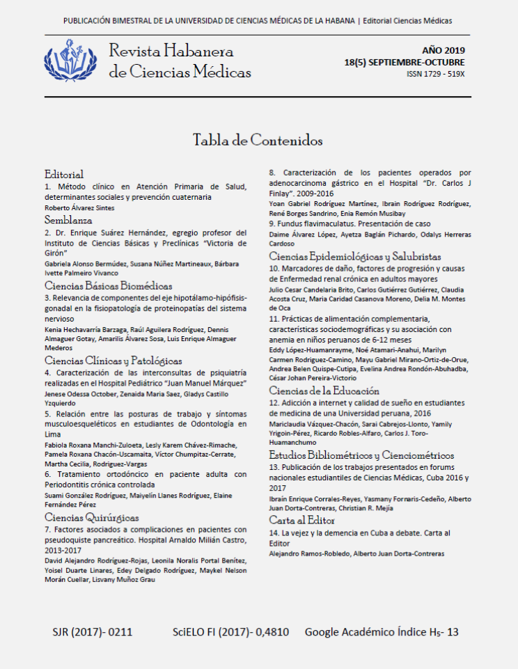
Fundus flavimaculatus. Presentación de caso
Palabras clave:
enfermedad de Stargardt, fundus flavimaculatus, distrofias maculares, tomografía de coherencia óptica, angiografía fluoresceínica, pronóstico.Resumen
Introducción: La Enfermedad de Stargardt, o fundus flavimaculatus es la distrofia macular juvenil más frecuente, responsable hasta 7 % de las distrofias maculares. Pueden ser difíciles de abordar por diversos motivos. El diagnóstico diferencial a veces es difícil, y varias de estas enfermedades producen ceguera legal a una edad relativamente joven.
Objetivo: Describir el proceso diagnóstico de una entidad poco común con forma de presentación infrecuente.
Presentación de caso: Se presenta el caso de una paciente de 21 años de edad, de raza blanca, con el diagnóstico de Enfermedad de Stargardt con fundus flavimaculatus. El diagnóstico se realizó teniendo en consideración los antecedentes patológicos personales, el examen físico mediante la oftalmoscopía directa e indirecta, la agudeza visual sin y con corrección, test de visión al color, angiografía fluoresceínica, tomografía de coherencia óptica (OCT) y electroretinograma. Se realizó una investigación de dicho tema por lo poco frecuente que resultan estas dos variantes de una misma enfermedad en la primera década de la vida.
Conclusiones: La mayoría de las distrofias retinianas tiene desde el punto de vista clínico sus semejanzas, en cambio su evolución y pronóstico pueden ser diferentes.Descargas
Citas
1. Medina López JP, Parada Vázquez RH, Rodríguez Gómez JA, Castillo Velázquez J. Enfermedad de Stargardt: presentación clínica de cuatro casos familiares. Oftalmol Clin Exp. 2016; 9(3):108-15.
2. Wright LS, Phillips MJ, Pinilla I, Hei D, Gamm DM. Induced pluripotent stem cells as custom therapeutics for retinal repair: progress and rationale. Exp Eye Res. 2014; 123:161-72.
3. Ritter M, Zotter S, Schmidt WM, Bittner RE, Deak GG, Pircher M, et al. Characterization of Stargardt disease using polarization-sensitive optical coherence tomography and fundus autofluorescence imaging. Invest Ophthalmol Vis Sci [Internet]. 2013;54(9):23. [Citado 20/09/2019]. Disponible en: https://iovs.arvojournals.org/article.aspx?articleid=2128276
4. Cai CX, Light JG, Handa JT. Quantifying the Rate of Ellipsoid Zone Loss in Stargardt Disease [Internet]. 2018;186:20. [Citado 20/09/2019]. Disponible en: https://www.clinicalkey.es/#!/content/playContent/1-s2.0-S0002939417304713?returnurl=https:%2F%2Flinkinghub.elsevier.com%2Fretrieve%2Fpii%2FS0002939417304713%3Fshowall%3Dtrue&referrer=https:%2F%2Fwww.researchgate.net%2F
5. Westeneng-van Haaften SC, Boon CJ, Cremers FP, Hoefsloot LH, den Hollander AI, Hoyng CB. Clinical and genetic characteristics of late-onset Stargardt’s disease. Ophthalmology [Internet]. 2012;119(6):[aprox. 22p.]. [Citado 20/09/2019]. Disponible en: https://www.clinicalkey.es/#!/content/playContent/1-s2.0-S0161642012000097?returnurl=https:%2F%2Flinkinghub.elsevier.com%2Fretrieve%2Fpii%2FS0161642012000097%3Fshowall%3Dtrue&referrer=https:%2F%2Fwww.ncbi.nlm.nih.gov%2F
6. McBain VA, Townend J, Lois N. Progression of retinal pigment epithelial atrophy in Stargardt disease. Am J Ophthalmol [Internet]. 2012;154(1):[aprox. 33p.]. [Citado 20/09/2019]. Disponible en: https://www.clinicalkey.es/#!/content/playContent/1-s2.0-S0002939412000700?returnurl=https:%2F%2Flinkinghub.elsevier.com%2Fretrieve%2Fpii%2FS0002939412000700%3Fshowall%3Dtrue&referrer=https:%2F%2Fwww.researchgate.net%2F
7. Wright LS, Phillips MJ, Pinilla I, Hei D, Gamm DM. Induced pluripotent stem cells as custom therapeutics for retinal repair: progress and rationale. Exp Eye Res. 2014; 123: 161-72.
8. Kanski JJ, Bowling B. Kanski Oftalmología clínica. 7ma. ed. Madrid: Elsevier; 2012.
9. Sangermano R, Khan M, Cornelis SS, Richelle V, Albert S, Garanto A, et al. ABCA4 midigenes reveal the full splice spectrum of all reported noncanonical splice site variants in Stargardt disease. Genome Res [Internet]. 2018;28(1):25. [Citado 20/09/2019]. Disponible en:https://www.ncbi.nlm.nih.gov/pmc/articles/PMC5749174/
10. Fujinami K, Lois N, Davidson AE, Mackay DS, Hogg CR, Stone EM,et al. A longitudinal study of Stargardt disease: clinical and electrophysiologic assessment, progression, and genotype correlations. Am J Ophthalmol [Internet]. 2013 Jun;155(6):23. [Citado 20/09/2019]. Disponible en: https://www.clinicalkey.es/#!/content/playContent/1-s2.0-S0002939413000743?returnurl=https:%2F%2Flinkinghub.elsevier.com%2Fretrieve%2Fpii%2FS0002939413000743%3Fshowall%3Dtrue&referrer=https:%2F%2Fwww.ncbi.nlm.nih.gov%2F
11. Testa F, Rossi S, Sodi A,Passerini I,Di Lorio V, Della Corte M,et al. Correlation between photoreceptor layer integrity and visual function in patients with Stargardt disease: implications for gene therapy. Invest Ophthalmol Vis Sci 2012; 53(8):4409-15.
12. Gérard X, Perrault I, Munnich A, Kaplan J, Rozet JM. Intravitreal Injection of Splice-switching Oligonucleotides to Manipulate Splicing in Retinal Cells. Mol Ther Nucleic Acids [Internet]. 2015 Sep;4(9):22. [Citado 20/09/2019]. Disponible en: https://www.ncbi.nlm.nih.gov/pmc/articles/PMC4877449/
13. Murray SF, Jazayeri A, Matthes MT, Yasumura D, Yang H, Peralta R, et al. Allele-specific inhibition of rhodopsin with an antisense oligonucleotide slows photoreceptor cell degeneration. Invest Ophthalmol Vis Sci [Internet]. 2015;56(11):25. [Citado 20/09/2019]. Disponible en: https://www.ncbi.nlm.nih.gov/pmc/articles/PMC5104522/
14. Lambertus S, Bax NM, Groenewoud JMM, et al. Asymmetric inter-eye progression in Stargardt disease. Invest Ophthalmol Vis Sci. 2016; 57(15):6824-30.
15. Cai CX, Locke KG, Ramachandran R, Birch DG, Hood DC. A comparison of progressive loss of the ellipsoid zone (EZ) band in autosomal dominant and x-linked retinitis pigmentosa. Invest Ophthalmol Vis Sci. 2014;55(11):7417-22.
16. Birch DG, Locke KG, Wen Y, Locke KI, Hoffman DR, Hood DC. Spectral-domain optical coherence tomography measures of outer segment layer progression in patients with x-linked retinitis pigmentosa. JAMA Ophthalmol. 2013; 131(9):1143-50.
17. Hariri AH, Velaga SB, Girach A, et al. Measurement and reproducibility of preserved ellipsoid zone area and preserved RPE area in a cohort of eyes with choroideremia. Am J Ophthalmol. 2017; 179:110-17.
18. Strauss RW, Ho A, Muñoz B, Cideciyan AV, Sahel JA, Sunness JS, et al. The Natural History of the Progression of Atrophy Secondary to Stargardt Disease (Prog- Star) studies: design and baseline characteristics: ProgStar Report No. 1. Ophthalmology. 2016; 123(4):817-28.
19. Cideciyan AV, Alemán TS, Swider M, Schwartz SB, Steinberg JD, Brucker AJ. Mutations in ABCA4 result in accumulation of lipofuscin before slowing of the retinoid cycle: a reappraisal of the human disease sequence. Hum Mol Genet. 2004; 13(5):525-34.


