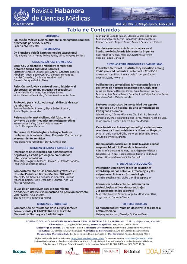
Protocol for direct vaginal cytology of laboratory rats
Keywords:
Estrous cycle, estrus, metestrus, diestrus, proestrus, direct vaginal cytology, Wistar rats.Abstract
Introduction: Direct vaginal cytology is a widely used method for the evaluation of the estrous cycle of laboratory rats, but the information about the procedures and interpretation of results is dispersed through literature, making its use difficult in investigations on reproduction.
Objective: To propose a protocol for performing direct vaginal cytology of laboratory rats as well as for the interpretation of the results.
Material and methods: The information obtained from literature and the experience of 10 years of investigation on reproduction in 250 female Wistar rats were combined following the established ethical principles. The procedures for obtaining samples by vaginal washing and by observation and analysis in humid state with light microscope equipped with digital camera were described.
Results: The main types of cells present in the vaginal washing and the characteristics that allow us to identify each phase or transitional phases of the estrus cycle are described. Aspects to take into consideration in the interpretation of the results, which include the association between hormonal changes, the cares in the obtaining of the sample and the influence of environmental factors, are discussed. Representative images and figures are included to illustrate the text.
Conclusions: The work consists of a study protocol of the estrus cycle of laboratory rats by direct vaginal cytology. It provides noninvasive, simple and cost-reducing procedures as well as essential knowledge for the interpretation of results which integrate a very useful guideline for experimental investigations on reproduction.
Downloads
References
1. Aritonanga TR, Rahayub S, Siraitc LI, Karod MB, Simanjuntake TP, Natzirf R, et al. The role of FSH, LH, estradiol and progesterone hormone on estrus cycle of female rats. IJSBAR [Internet]. 2017 [Citado 19/11/2020];35(1):92-100. Disponible en: https://www.gssrr.org/index.php/JournalOfBasicAndApplied/article/view/7834
2. Folorunsho A, Eghoghosoa R. Staging of the estrous cycle and induction of estrus in experimental rodents: an update. Fertil Res Pract [Internet]. 2020 [Citado 19/11/2020];6(5):1-15. Disponible en: http://doi.org/10.1186/s40738-020-00074-3
3. Karwasik Kajszczarek K, Chmiel Perzyńska I, Marcin J, Billewicz Kraczkowska A, Pedrycz A, Smoleń A, et al. Impact of experimental diabetes and chronic hypoxia on rat fetal body weight. Ginekol Pol [Internet]. 2018 [Citado 19/11/2020]; 89(1):20-4. Disponible en: http://doi.org/10.5603/GP.a2018.0004
4. Essiet GA, Akuodor GC, Oj D, Nwokike MO, Eke DO, Chukwumobi AN. Effects of Salacia lehmbachii ethanol root bark extract on estrous cycle and sex hormones of female albino rats. Asian Pac J Reprod [Internet]. 2018 [Citado 19/11/2020]; 7(6):274-9. Disponible en: http://doi.org/10.4103/2305-0500.246347
5. Alonso Caraballoa Y, Ferrario CR. Effects of the estrous cycle and ovarian hormones on cuetriggered motivation and intrinsic excitability of medium spiny neurons in the Nucleus Accumbens core of female rats. Horm Behav [Internet]. 2019 [Citado 19/11/2020];116:104583. Disponible en: http://doi.org/10.1016/j.yhbeh.2019.104583
6. Shi Z, Wong J, Brooks VL. Obesity: sex and sympathetics. Biology of Sex Differences [Internet]. 2020 [Citado 19/11/2020];11(10):1-13. Disponible en: http://doi.org/10.1186/s13293-020-00286-8
7. Chari T, Griswold S, Andrews NA, Fagiolini M. The stage of the estrus cycle is critical for interpretation of female mouse social interaction behavior. Front Behav Neurosci [Internet]. 2020 [Citado 19/11/2020];14:113. Disponible en: http://doi.org/10.3389/fnbeh.2020.00113
8. Jafarey R, Jaffri SA. Study on the estrous cycle regularity of cryopreserved rat ovarian tissues after heterotopic transplantation. Open Journal of Obstetrics and Gynecology [Internet]. 2016 [Citado 19/11/2020];6:293-8. Disponible en: http://doi.org/10.4236/ojog.2016.65037
9. Cora MC, Kooistra L, Travlos G. Vaginal cytology of the laboratory rat and mouse: review and criteria for the staging of the estrous cycle using stained vaginal smears. Toxicol Pathol [Internet]. 2015 [Citado 19/11/2020];43(6):776-93. Disponible en: http://doi.org/10.1177/0192623315570339
10. Mohammed S, Sundaram V. Comparative study of metachromatic staining methods in assessing the exfoliative cell types during oestrous cycle in sprague-dawley laboratory rats. Int J Morphol [Internet]. 2018 [Citado 19/11/2020];36(3):962-8. Disponible en: http://doi.org/10.4067/S0717-95022018000300962
11. Pantier LK, Li J, Christian CA. Estrous cycle monitoring in mice with rapid data visualization and analysis. Bio-protocol [Internet]. 2019 [Citado 19/11/2020]; 9(17):e3354. Disponible en: http://doi.org/10.21769/BioProtoc.3354
12. Committee for the Update of the Guide for the Care and Use of Laboratory Animals. Guide for the Care and Use of Laboratory Animals. 8 ed [Internet]. Washington: National Academies Press; 2011. [Citado 19/11/2020]. Disponible en: http://doi.org/10.17226/12910
13. Fernández T, Clapés S, Suárez G, González L, Rodríguez S, García MA. Alpha-tocopherol supplementation diminishes the renal damage caused by experimental diabetes. J Diabetes Metab [Internet]. 2015 [Citado 19/11/2020];6(1):1000478. Disponible en: http://doi.org/10.4172/2155-6156.1000478
14. Christensen A, Bentley GE, Cabrera R, Ortega HH, Perfito N, Wu TJ, et al. Hormonal Regulation of Female Reproduction. Horm Metab Res [Internet]. 2012 [Citado 19/11/2020];44(8):587-91. Disponible en: http://doi.org/10.1055/s-0032-1306301
15. Hill JW, Elias CF. Neuroanatomical framework of the metabolic control of reproduction. Physiol Rev [Internet]. 2018 [Citado 19/11/2020];98(4):2349-80. Disponible en: http://doi.org/10.1152/physrev.00033.2017
16. Sanabria V, Bittencourt S, de la Rosa T, Livramento J, Tengan C, Scorza CA, et al. Characterization of the estrous cycle in the amazon spiny rat (Proechimys guyannensis). Heliyon [Internet]. 2019 [Citado 19/11/2020];5(12):e03007. Disponible en: http://doi.org/10.1016/j.heliyon.2019.e03007
17. Srinivasan MR, Sabarinathan A, Geetha A, Shalini K, Sowmiya M. A comparative study on staining techniques for vaginal exfoliative cytology of rat. J of Pharmacol & Clin Res [Internet]. 2017 [Citado 19/11/2020];3(3):1-5. Disponible en: http://doi.org/10.19080/JPCR.2017.03.555615
18. Yaw AM, McLane Svoboda AK, Hoffmann HM. Shiftwork and light at night negatively impact molecular and endocrine timekeeping in the female reproductive axis in humans and rodents. Int J Mol Sci [Internet]. 2021[Citado 19/11/2020];22(1):324. Disponible en: http://doi.org/10.3390/ijms22010324
19. An G, Chen X, Li C, Zhang L, Wei M, Chen J, et al. Pathophysiological changes in female rats with estrous cycle disorder induced by long-term heat stress. Biomed Res Int [Internet]. 2020 [Citado 19/11/2020];2020:1-10. Disponible en: http://doi.org/10.1155/2020/4701563
20. Sampath G. Comparative study on the estimation of estrous cycle in mice by visual and vaginal lavage method. J Clin Diagnostic Res [Internet]. 2017 [Citado 19/11/2020];11(1):AC05-AC07. Disponible en: http://doi.org/10.7860/JCDR/2017/23977.9148


